From Texas Public Radio:
Each year in the U.S., as many as 3,000 children drown in water. About two-thirds of them are resuscitated, but the brain damage is usually devastating.
Now, a newly-published study by a San Antonio researcher shows there may come a day when doctors can target the brain damage from drowning and perhaps even treat it in the emergency room to change the outcome.
Summer time is prime time for swimming. Water lures even the youngest of children and can lead to disaster.
San Antonio mother Liz Tullis knows this the hard way. Her son Conrad was visiting at his grandfather’s when an accident happened. “When Conrad was 17 months old, he fell into a swimming pool,” she says.
Then, the unthinkable. Her son was breathing, but he wasn’t moving. He was fed through a tube. She had to decide whether to withdraw treatment, put him in long-term care, or take him home.
“There’s not really a road map,” Tullis says.
She took Conrad home. That was 13 years ago. During that time, her son started waking and sleeping, and reacting, she thought, to what was going on around him. He is awake and aware, but unable to communicate.
She was convinced it was no coincidence.
“I started sensing he understood what was going on,” Tullis says.
This determined and curious mother sought out help from a UT Health San Antonio neurologist, Peter Fox, and his scanning technology.
“The brain is the organ that’s most greedy for oxygen,” Fox says.
Fox says when a child inhales water and drowns, their heart stops beating very quickly. By the time they get to an emergency room, they’re usually comatose or convulsing.
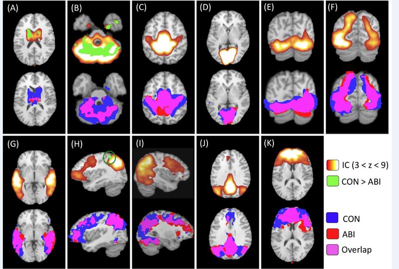
Using MRI machines at the Research Imaging Institute, Fox scanned Conrad and nine other children from around the country in similar condition. What he found was surprising.
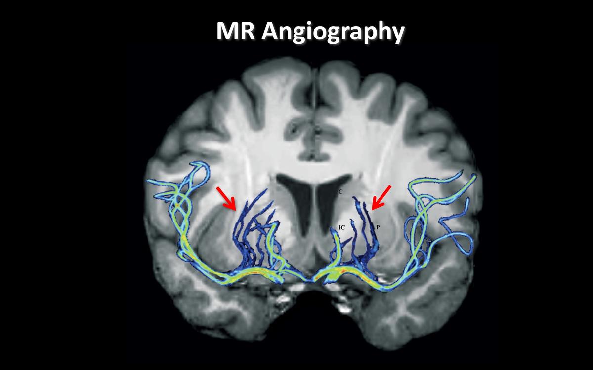
“The lesion is smack in the motor fibers,” he says.
Using tests that can reveal both structural damage and functional capabilities, Fox has now published his work, which shows the damage in children whose brains have been starved of oxygen in a drowning, is concentrated in a small, specific area in the basal ganglia found deep and near the center of the brain.


