This story originally appeared on Houston Public Media.
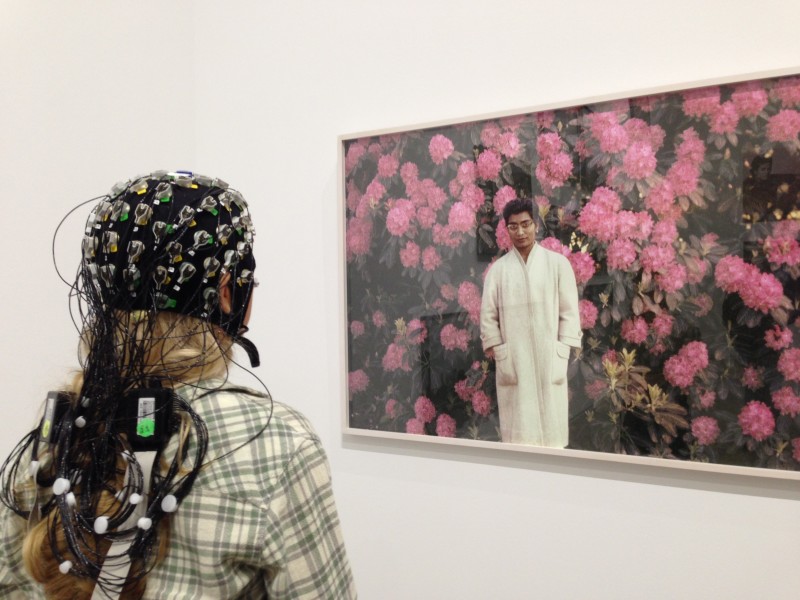
In the lobby of the Blaffer Art Museum at the University of Houston, I’m in the process of becoming a living Medusa. Squeezed over my head is a tightly-fitted cap, resembling the kind you see swimmers wear. Dangling from it are 64 wires, each attached to an electrode. They’ll monitor what’s going on up there as I stroll around the museum gallery and check out the art.
Research assistants Anastasiya Kopteva and Andrew Paek are in the process of applying gel to my head to make the electrodes stick. At last, all the lights on the cap have turned green, meaning we have contact.
A tablet computer showing my brain waves in little squiggly lines hangs around my neck, accessorized with a fanny pack holding the various other tech gear. The ensemble is complete with a small camera clipped to the front of my shirt. That’ll let researchers know what art piece I’m looking at when they go back to examine the results.
“So, what’s next?” I ask Kopteva.
“Let’s view art,” she answers.
And through the museum I go, followed closely by Kopteva and Paek, who are pushing a cart with a laptop behind me. I ask Paek what’s up with the laptop.
“We get electricity from your brain, we send it wireless, (and) our computer picks it up,” he said.
At one point, I stop to check my tablet and ask Paek if there’s any way to draw conclusions from the data so far. Can we notice any trends?
“Um, no,” he laughs. Apparently, it’s a little more complicated than that.
Fast forward two weeks.
Getting the EEG results involves a trip over to Dr. Jose Luis Contreras-Vidal’s lab inside the University of Houston Engineering School. At a large square table, we’re staring a screen on the wall, which has images of my 64 EEG channels projected onto it.
But at this point, making sense of exactly what all those squiggly lines mean is an intricate process.
“And so what we do first is clean the signal from anything that’s not related to the task,” Contreras-Vidal explains. “That includes eye blinks and eye movements.”
Which, in turn, involves the tedious task of combing through the data and removing the unneeded material, referred to as “artifacts.” Once those artifacts are removed, they’re left with pure brain activity.
So what about that flickering projector installation piece that I had a particular attachment to? What was my brain doing at that moment?
“That’s the visual cortex,” Contreras-Vidal says, pointing to a red section at the back of my head. “So you see that there’s a bump in the bottom plot around 11 hertz. That means the brain activity of your visual cortex is oscillating around 11 hertz.”
Which doesn’t tell us a whole lot – yet. But when researchers start looking at data from hundreds of people – then thousands – they start to notice trends.
“We know that that part of your brain is active and we want to see how that activation relates to other areas in the brain,” he says. “How the information travels and how long it takes to get there is important.”
One of his hopes is to inform the clinical neuroscience field about how best to use art to treat neurological and mental disorders. It’s a long-term project uniting fields that aren’t often linked.
“This is really work that’s at the interface between the arts, science and engineering. Studying creativity, studying how the brain perceives art – and how we can use that to push science and engineering,” he said.
Next on the horizon is an international conference this summer in Cancun. It’ll include some of the world’s thought-leaders in neuroscience, engineering, education, and medicine.


